
Disease Diagnosis
Many diseases we come across in pets require laboratory investigations and other diagnostic testing for us to reach a diagnosis. Tests such as blood and urine tests, xrays, ultrasound and many more give us valuable information about the function of certain organs and body systems in the face of illness that allow us to reach a diagnosis and therefore be able to offer the most appropriate treatment. The following outlines the tests we are able to offer here at DMVS, and how we use them in the diagnostic procedure.
Physical Examination
Every diagnostic process starts with a thorough physical examination. A thorough examination of your pet in the face of an illness can give us clues as to where in the body the problem lies. This is of particular importance in illnesses that are more vague in nature, and may often need to be repeated a number of times over a couple of days, each time potentially gaining more information.
For example, a dog with frequent urination and a painful abdomen may have a urinary tract infection, bladder stones, a bladder tumour (cancer), or a number of other diseases, but most likely the disease would be localised to the urinary tract. From our physical examination, we can then focus our initial diagnostic testing on this body system.
Haematology & Biochemistry

This is a general ‘blood test’. Haematology refers to the red and white blood cells (i.e. the cellular component of blood), and biochemistry refers to many markers of different organs, as well as the electrolyte and acid/base balance of the body, that are present in the liquid component of the blood. The information gained from this general blood test is enormous, as it covers many major organs and in many cases, can indicate mild vs severe disease.
After a physical examination, haematology & biochemistry would have to be our next most frequent form of diagnostic testing. This is due to it’s broad range of diseases it can give us information about – most systems in the body! Additionally, it is quick and easy to perform (we run the bloods in-house), we get results quickly, it is well tolerated by most pets, and best of all, we gain a lot of information across a broad range of systems from this one test.
For example, a case of a vomiting dog may be simply a mild gut upset from eating the wrong food, or it could involve a malfunction in a major organ like the liver, kidneys or pancreas. If our veterinary physical examination of the dog reveals only mild change to the pet, we would try 24 hours off food with water available, then a bland diet. Some unwell pets will recover. Other more seriously affected pets have significant signs of disease on examination or do not respond to the above treatment and continue to vomit. These pets will need blood tests to help localise the organ/s affected. We are then equipped with knowledge to treat the specific disease. Without that information, some diseases advance and can potentially prevent a full recovery. Our blood tests allow to determine the cause of some diseases early, allowing appropriate treatment to be given earlier, and with this there is generally better results and outcomes.
Specialised Biochemistry
We also have more specialised biochemistry testing available in-house, which are used for both disease diagnosis and monitoring of chronic disease. Examples of these include a canine specific pancreatic lipase test (specific test for canine pancreatitis), cortisol testing (for diagnosis of hyperadrenocorticism or hypoadrenocorticism, and the monitoring of these diseases) and total T4 measurement (for diagnosis and monitoring of hyperthyroidism and hypothyroidism). This aids in rapid diagnosis and convenient monitoring of these diseases.
Clotting tests
We run both a basic activated clotting test and a more specific PT / aPTT clotting test in house. These help us to rapidly diagnose clotting disorders such as ratbait toxicity and snake envenomations to allow timely emergency treatment. Once the clotting disorder has been treated, we often run follow-up tests to ensure everything has gone back to normal when medication has finished.
Urinalysis
This is a ‘urine test’. A urinalysis involves a dipstick examination (which looks at various components such as glucose, blood and white blood cells), measurement of urine specific gravity (a measurement of the concentration of the urine), and a sediment examination (a view of the microscopic components of the urine such as bacteria, blood, white blood cells and urine crystals).
We often perform a urinalysis in conjunction with haematology & biochemistry to further determine the nature of diseases. For example, let’s say the vomiting dog described above is also dehydrated. The blood test may show elevated kidney enzymes, indicating possible kidney disease, severe dehydration (which would also be noted on their physical examination) or possibly even obstruction of the urinary tract. If we perform a urinalysis on this patient, the concentration of the urine will tell us if the animal is dehydrated (very concentrated urine) or has severe kidney disease (the urine will not be concentrated because the kidneys are not performing their function to maintain water balance). Diagnosis of urinary obstruction is a little different, but a urinalysis would still be performed in these patients to try to find the cause of the obstruction!
With this knowledge we can again give more targeted treatment and give more information regarding prognosis for return to good health.
Faecal floatation
A faecal floatation is where we mix some faeces with very highly concentrated salt water so that worm eggs and other organisms float to the surface of the salt water. We catch them on a slide and examine this under the microscope. We can see worm eggs, giardia and coccidia. This can aid in the diagnosis of the possible cause of diarrhoea, usually when our regular treatments don’t resolve the problem.
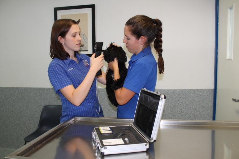
Ear cytology (Ear smear)
There are many people who have been through the frustration of ear infections, and worse still, recurrent ear infections! You will all know the importance of ear cytology right from the beginning to ensure the best possible chance of successful treatment.
Ear testing first involves looking down the ear with an otoscope to examine the ear canal and eardrum. This can find nasties like grass seeds and other foreign bodies, or can show an inflamed ear canal, or even a ruptured eardrum. When an ear looks infected, we often take a swab of the discharge and examine it under the microscope to identify the nature of the cause, whether it be bacteria or yeast, or a mixture of the two. This then allows us to select the most appropriate medication to give the best possible outcome. Ear infections can often be challenging, so we need to repeat this at the end of the course of medication to ensure the infection has cleared.
Skin Testing
Skin testing is indicated in many cases of itchy dogs, lumps and bumps, as well as unusual diseases such as auto-immune skin conditions. We can perform superficial and deep skin scrapings in-house to detect various forms of mites. We also perform sticky tape preps (yep, as silly as it sounds!) of skin to detect superficial bacterial and yeast infections of the skin. More
Histopathology
Histopathology involves the evaluation of a sample of tissue under a microscope by a veterinary pathologist. This is done at an external laboratory, and includes an opinion on the case from a specialist veterinary pathologist. The tissue sampled could be a lump that has been removed, a skin biopsy, or a biopsy of another organ, such as liver or intestines. This evaluation of tissues by a pathologist can give us such valuable information, and in most cases, can give us a definitive diagnosis for a patient. Therapy for that patient can then be tailored according to the specific diagnosis. In the case of lumps that have been removed, we get a diagnosis of the cell type involved, and therefore can predict the behaviour of the lump for the future. We also assess the margins of the lump to see if it has been completely removed down to the microscopic level. Histopathology has many functions in our clinic, and always gives us helpful information when performed.
Radiography (X-rays)

X-rays are a very helpful imaging tool in such a wide range of applications. We use x-rays for anything from hip scoring in breeds such as Labradors and German Shepherds, right through to cases of vomiting and diarrhoea, broken bones, heart disease, ingestion of a foreign body, and the list just goes on and on and on. X-ray images show us bone and soft tissue and can identify broad changes in the structures that are imaged.
This x-ray is of a dog that is just about to have puppies! We sometimes x-ray expectant mothers just prior to their birth to predict the number of pups to expect to ease a little pressure on us on the big day! It is also very useful if there is any question as to whether there are any pups left to go! See if you can count how many there are here - it gets a bite tricky with the big litters like this one! Hint - count the rounded skulls!
Ultrasound

Most people will be familiar with this great non-invasive diagnostic imaging tool. Ultrasound can be performed on many parts of the body in order to visualise internal organs. We most commonly use ultrasound in our clinic for disorders involving the abdomen and heart. We can evaluate the size and shape of many different organs, and detect some abnormalities in structure of these organs. Abdominal tumours (cancers) can also often be found on ultrasound. On a brighter note, we also diagnose pregnancy in dogs and cats using ultrasound.
Electrocardiogram (ECG) and blood pressure measurement

An Electocardiogram is used to detect and diagnose heart arrhythmias
For a complete cardiac workup, a combination of ultrasound, radiography, ECG and blood pressure studies can be performed. The prognosis can be more accurately gauged by using all or a combination of these tools.
A normal heart beat produces an electric signal through the heart muscle to stimulate it to beat. This is a heavily coordinated signal, producing a coordinated contraction of the heart muscle. Along with the heart valves, this coordinated contraction allows blood to flow efficiently around the body. In certain heart disease (and some other medical conditions), this system is disrupted and the electrical current running through the heart muscle is disrupted. This can be detected using electrocardiography. Electrocardiography (ECG) is used in our clinic in the case of abnormal heart rhythms (cardiac arrhythmias). This may be that the heart is beating too fast, too slow or irregularly. This can be important in a range of medical conditions, not just those primarily relating to the heart. Understanding the changes of the electrical pattern of the heart can lead to better therapy for each specific heart condition.
Blood pressure is vital for blood to continue to circulate around the body. It is important to measure blood pressure in a huge range of situations. Dogs and cats can suffer from high blood pressure (hypertension) just like people do. Medication for hypertension can lengthen the lives of the pets that suffer from it. Other times we use blood pressure measurement include anaesthesia and surgery, trauma and as part of monitoring for kidney disease. Blood pressure measurement is often another piece of useful information along with a whole lot more that allows us to provide the best care possible for pets.
Tonometry
Tonometry is to measure intraocular pressure, or IOP (the pressure inside the eyeball). You may have had this performed at the optometrist before. The IOP is important for us to measure in cases of glaucoma (high IOP) and cases of uveitis (low IOP). Glaucoma is a very painful condition of the eye that can come on very suddenly and if not diagnosed and treated immediately, can cause permanent vision loss. Tonometry is obviously of great importance in the diagnosis, and then the management of these cases, to ensure that medication is keeping the IOP within the normal range.
Uveitis is inflammation of the vascular portion of the eye, including the iris (the coloured part). This can occur due to trauma, infection or auto-immune inflammation, as well as other causes. The low pressure can cause problems to the eye, so medication is required to lift the IOP back up into normal range. Again, having tonometry available allows us to provide medication to these patients in order to have the best possible management of the changes to their eyes.
Endoscopy

Endoscopy refers to the process of using a long, thin, flexible camera to visualise inside certain structures, usually hollow organs or a body cavity. This can be helpful in the diagnostic process as this allows us to directly visualise internal structures such as the inner surface of the oesophagus, inside the trachea (windpipe) and in the nasal cavity. It is a great tool to remove things such as grass seeds that manage to get up noses and down the windpipe, and also to visualise things such as ulcers in the oesophagus. It is another great non-invasive diagnostic tool that helps us out in a lot of tricky cases.